39 cell wall diagram with labels
Animal Cell Diagram with Label and Explanation: Cell Structure, Functions Diagram of Animal Cell Below is the diagram of the animal cell which shows the organelles present in it. The cell is covered with cytoplasm which consists of cell organelles in it. The nucleus is covered with a rough Endoplasmic Reticulum and other organelles each designed for a specific purpose. › cells › bactcellInteractive Bacteria Cell Model - CELLS alive In the space are enzymes and other proteins that help digest and move nutrients into the cell. Cell Wall: Composed of peptidoglycan (polysaccharides + protein), the cell wall maintains the overall shape of a bacterial cell. The three primary shapes in bacteria are coccus (spherical), bacillus (rod-shaped) and spirillum (spiral).
PDF Plant Cell Diagram - Edraw Soft Plant Cell Golgi vesicles Golgi apparatus Ribosome Smooth ER(no ribosomes) Nucleolus Nucleus Rough ER(endoplasmic reticulum) Large central vacuole Amyloplast(star ch grain) Cell wall Cell membrane Chloroplast Vacuole membrane Raphide crystal Mitochondrion Druse crystal
Cell wall diagram with labels
Animal Cell Labeled Detailed / Plant And Animal Cell Diagram Labeled ... Animal Cell Labeled Detailed / Plant And Animal Cell Diagram Labeled Drawing Free Image Download : Also know that the membrane is not a rigid cell wall like in plant cells.. Draw a labelled diagram of a animal cell and plant cell, images of animal cell, images of plant cell. 4.2 differences between plant cell and animal cell plant cell 1 ... Plant Cells: Labelled Diagram, Definitions, and Structure The cell wall is made of cellulose and lignin, which are strong and tough compounds. Plant Cells Labelled Plastids and Chloroplasts Plants make their own food through photosynthesis. Plant cells have plastids, which animal cells don't. Plastids are organelles used to make and store needed compounds. Chloroplasts are the most important of plastids. Spirogyra Labelled Diagram Draw a neat diagram of Spirogyra and label the following parts: i. Outermost layer of the cell. ii. Organelle that performs the function of. Each cell of Spirogyra filament is cylindrical and consists of 2 parts: cell wall and protoplast. The cell wall surrounds the protoplast, is protective and consists of.
Cell wall diagram with labels. PDF Plant Cells - Definition, Diagram, Structure & Function - NFEI Plant Cells - Definition, Diagram, Structure & Function The cell is the basic unit of life in all organisms. Like humans and animals, plants are also composed of several cells. The plant cell is surrounded by a cell wall which is involved in providing shape to the plant cell. Apart from the cell wall, there are other organelles that are Label the cell - Teaching resources G5 Science. Label the Plant Cell Labelled diagram. by Koneal2. G7 Biology. Label Animal Cell Organelles Labelled diagram. by Britter. Correctly Label the Bacteria (Prokaryotic) Cell Labelled diagram. by Bronwyn12. Label Plant and Animal Cell Labelled diagram. sciencequiz.net › newjcscience › jcbiologyThe Cell - ScienceQuiz.net The diagram shows a plant cell as seen under a microscope. Two of the labels are incorrect. What are they? ... A is the cell wall and DNA is located inside B.? Bacteria shapes, structure and diagram - Jotscroll The bacteria shapes, structure, and labeled diagrams are discussed below. Sizes The sizes of bacteria cells that can infect human beings range from 0.1 to 10 micrometers. Some larger types of bacteria such as the rickettsias, mycoplasmas, and chlamydias have similar sizes as the largest types of viruses, the poxviruses.
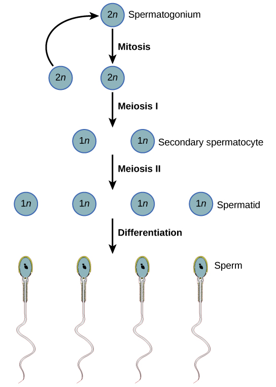
Plant and Animal Cell: Labeled Diagram, Structure, Function - Embibe Cell Wall: 1. Non-living, rigid, outer boundary. 2. Made up of cellulose, hemicellulose, pectin, lignin, etc. 3. There are many layers, like the middle layer, primary cell wall in a typical plant cell wall. 4. Fungal cell wall is made up of chitin (not cellulose). 5. Protective and provide shape and size. 6. Found only in plant cells. Plasma ... Elodea Leaf Cell Diagram Elodea Leaf Cell Diagram. The Elodea leaf is composed of two layers of cells. Only one layer of cells is in focus when using the high. Examining elodea (pondweed) under a compound microscope. solution) and a coverslip and observe the chloroplasts (green structures) and the cell walls. In this lesson, students microscopically observe various ... Meiosis In Plant Cell Diagram Labeled : Functions and Diagram Meiosis is the division of a germ cell into four sex cells (e.g. egg or sperm), each with half the number of chromosomes of the parent cell. Meiosis is a form of nuclear division that results in the production of haploid cells from diploid cells; It produces gametes in plants and animals that are used in sexual reproduction; It has many ... › basic-cells › label-diagramLabel Cell Parts | Plant & Animal Cell Activity | StoryboardThat Have your students label a plant and animal cell using one of the landscape poster layouts (small or large). Students will create a cell diagram labeled with the different organelles of plant and animal cells. The cell diagrams are easily colorable, allowing students to differentiate the different parts of the plant and animal cell quickly.
PDF PLANT CELL DIAGRAM - abcteach.com for the cell. K. The "control center" of the cell; this contains the cell's DNA. L. A thick, stiff membrane that surrounds the plant cell and supports the plant structure. M. A thin, semi-permeable membrane that surrounds the cell, inside the cell wall. N. An organelle that stores molecules such as starch and pigment. __ 1. Nucleus __ 2. Structure of Fungal Cell (With Diagram) | Fungi The living substance of the cell within the cell wall is the protoplast. It lacks the chloroplasts but is differentiated into the other usual cell parts such as plasma or cell membrane, vacuolated cytoplasm, cell organelles and one or more nuclei. Cell Membrane: It is a delicate, extremely thin, living membrane which closely invests the protoplast. en.wikipedia.org › wiki › NephronNephron - Wikipedia The nephron uses four mechanisms to convert blood into urine: filtration, reabsorption, secretion, and excretion.: 395–396 These apply to numerous substances. The structure and function of the epithelial cells lining the lumen change during the course of the nephron, and have segments named by their location and which reflects their different functions. Cell Organelles- Definition, Structure, Functions, Diagram In a plant cell, the cell wall is made up of cellulose, hemicellulose, and proteins while in a fungal cell, it is composed of chitin. A cell wall is multilayered with a middle lamina, a primary cell wall, and a secondary cell wall. The middle lamina contains polysaccharides that provide adhesion and allow binding of the cells to one another.
Human Reproductive Anatomy and Gametogenesis | Boundless Biology
Animal Cell Diagram Vacuole Labeled : Functions and Diagram Vacuoles are a kind of microscopic cellular structure named organelle. In animal cells, they are small and typically transport materials into and out of the cell. Vacuoles serve many functions, depending on the needs of the cell. One of the most intricate tasks that healthiness experts face throughout their interplay with patients is helping ...

The Plant Cell
Plant Cell: Diagram, Types and Functions - Embibe Exams Plant Cell Wall It is a rigid layer that is composed of cellulose, glycoproteins, lignin, pectin and hemicellulose. It is located outside the cell membrane and is completely permeable. The primary function of a plant cell wall is to protect the cell against mechanical stress and to provide a definite form and structure to the cell.
Free Heart Diagram Unlabeled, Download Free Heart Diagram Unlabeled png images, Free ClipArts on ...
Animal Cell Labelling Activity | Primary Resources | Twinkl This Animal Cell Labelling Activity is the perfect way to help children consolidate their learning on the biology of cells. Cells are the building blocks of life, and they're important for all budding young biologists to understand. With that in mind, we have created a range of resources to help those who teach teach their students about cells:

Index of /kvhs/biology
› 2018Origin 2018 Feature Highlights Directly type unicode characters into header rows such as Long Name or comments. Type the code and press ALT+X to insert the desired character, or right-click in edit mode to open Character Map to select character for insertion, Unicode characters will display everywhere including graph labels and legends, and also in dialog such as Object Manager, Layer Contents, and Plot Details.
Post a Comment for "39 cell wall diagram with labels"