38 the human eye without labels
Human Eye Anatomy - Parts of the Eye and Structure of the ... - Health Jade The eye is a hollow, spherical structure about 2.5 centimeters in diameter. Its wall has three distinct layers—an outer (fibrous) layer, a middle (vascular) layer, and an inner (nervous) layer. The spaces within the eye are filled with fluids that help maintain its shape. Figure 6. Structure of the human eye. human internal anatomy Eye anatomy human muscles eyeball front clipart labels without muscle etc usf edu clipground illustration clip tiff craft. Human body with internal organs, nervous system (print #13011047) ... eye anatomy human muscles eyeball front clipart labels without muscle etc usf edu clipground illustration clip tiff craft. Skeleton, Bones And Internal ...
Eye Anatomy: Parts of the Eye and How We See Behind the anterior chamber is the eye's iris (the colored part of the eye) and the dark hole in the middle called the pupil. Muscles in the iris dilate (widen) or constrict (narrow) the pupil to control the amount of light reaching the back of the eye. Directly behind the pupil sits the lens. The lens focuses light toward the back of the eye.

The human eye without labels
Cornea of the Eye - Definition and Detailed Illustration Cornea Definition. The cornea is the clear front surface of the eye. It lies directly in front of the iris and pupil, and it allows light to enter the eye. Viewed from the front of the eye, the cornea appears slightly wider than it is tall. This is because the sclera (the "white" of the eye) slightly overlaps the top and bottom of the anterior ... PDF Parts of the Eye - National Institutes of Health Eye Diagram Handout Author: National Eye Health Education Program of the National Eye Institute, National Institutes of Health Subject: Handout illustrating parts of the eye Keywords: parts of the eye, eye diagram, vitreous gel, iris, cornea, pupil, lens, optic nerve, macula, retina Created Date: 12/16/2011 12:39:09 PM Eye anatomy: A closer look at the parts of the eye In a number of ways, the human eye works much like a digital camera: Light is focused primarily by the cornea — the clear front surface of the eye, which acts like a camera lens. The iris of the eye functions like the diaphragm of a camera, controlling the amount of light reaching the back of the eye by automatically adjusting the size of the ...
The human eye without labels. Anatomy of the Eye | Kellogg Eye Center | Michigan Medicine Anatomy of the Eye. Choroid. Layer containing blood vessels that lines the back of the eye and is located between the retina (the inner light-sensitive layer) and the sclera (the outer white eye wall). Ciliary Body. Structure containing muscle and is located behind the iris, which focuses the lens. Cornea. Human eye - Wikipedia The human eye is a sensory organ, part of the sensory nervous system, that reacts to visible light and allows us to use visual information for various purposes including seeing things, keeping our balance, and maintaining circadian rhythm . The eye can be considered as a living optical device. Category:Human eyes - Wikimedia Commons Pages in category "Human eyes" This category contains only the following page. H. Human eye; Media in category "Human eyes" The following 179 files are in this category, out of 179 total. 2016-09-21 Herstmonceux Observatorium 06.jpg. Afghani eye.png 118 × 106; 21 KB. Alfred Hitchcock's The Wrong Man trailer 02.png. An is always on you.jpg. The Eyes (Human Anatomy): Diagram, Optic Nerve, Iris, Cornea ... - WebMD The weaker eye, which may or may not wander, is called the "lazy eye." Astigmatism: A problem with the curve of your cornea. If you have it, your eye can't focus light onto the retina the way it...
File:Diagram of human eye without labels.svg - Wikimedia Size of this PNG preview of this SVG file: 410 × 430 pixels. Other resolutions: 229 × 240 pixels | 458 × 480 pixels | 732 × 768 pixels | 976 × 1,024 pixels | 1,953 × 2,048 pixels. Original file (SVG file, nominally 410 × 430 pixels, file size: 277 KB) File information. Structured data. ear diagram without labels ear diagram without labels Label Parts of the Human Ear we have 9 Images about Label Parts of the Human Ear like Ear Labeling Quiz - Human Anatomy, Label Parts of the Human Ear and also 10 Best Images of Label Ear Diagram Worksheet - Blank Rock Cycle. Here it is: Label Parts Of The Human Ear academic.udayton.edu Anatomy of the eye: Quizzes and diagrams - Kenhub Here you can see all of the main structures in this area. Spend some time reviewing the name and location of each one, then try to label the eye yourself - without peeking! - using the eye diagram (blank) below. Unlabeled diagram of the eye Click below to download our free unlabeled diagram of the eye. Quiz: Label The Parts Of The Eye - ProProfs How much did you get to understand about the human eye? Take up this quiz and find out! Questions and Answers. 1. A is pointing to what part of the eye? A. Cornea. B. Optic Nerve.
eyeball diagram to label; labels without eye hypothalamus thalamus clipart front brain label parts stem cerebellum clipground. Label The Eye . eye diagram blank human labeled anatomy label eyeball worksheet quiz drawing labels purposegames answers parts printable ear eyes activities coloring. Human Eye Diagram, How The Eye Work -15 Amazing Facts Of Eye Anatomy of the Eye | Johns Hopkins Medicine Ciliary body. The part of the eye that produces aqueous humor. Cornea. The clear, dome-shaped surface that covers the front of the eye. Iris. The colored part of the eye. The iris is partly responsible for regulating the amount of light permitted to enter the eye. Lens (also called crystalline lens). Activity Sheet 1: How the Eyes Work | Human eye diagram, Teaching ... Description Use these simple eye diagrams to help students learn about the human eye. Three differentiated worksheets are included: 1. Write the words using a word bank 2. Cut and paste the words 3. Write the words without a word bank Labels include: eyebrow, eyelid, eyelashes, pupil, iris, and sclera. Eye Diagram Teaching Resources | Teachers Pay Teachers The Human Eye Overview Reading Comprehension and Diagram Worksheet. by. Teaching to the Middle. 63. $1.50. Zip. This passage briefly describes the human eye (900-1000 Lexile). 14 questions (matching and multiple choice) assess students' understanding. Students label a diagram of 6 parts of the eye. I've included a color and BW version, as well ...
yog-blogsoth: THE SKINLESS ONE (NYARLATHOTEP)
Eye Anatomy: 16 Parts of the Eye & Their Functions The lens of the eye (or crystalline lens) is the transparent lentil-shaped structure inside your eye. This is the natural lens. It is located behind the iris and to the front of the vitreous humor (vitreous body). The vitreous humor is a clear, colorless, gelatinous mass that fills the gap between the lens and the retina in the eye.

Notes of Ch 11 Human Eye and Colourful World| Class 10th Science « Study Rankers
Human Eye Explorer Even microscopic structures, usually not visible for the human eye, can be explored: see inside the retina or cornea and discover its layers and cells from all perspectives. Additional functions like the presentation editor, the media library or labeling and color tools, make the Human Eye Explorer a unique software.

Eye Diagram Without Labels | via Anatomy Pictures Gallery if… | Flickr
The Human Eye | Boundless Physics | | Course Hero The human eye is the gateway to one of our five senses. The human eye is an organ that reacts with light. It allows light perception, color vision and depth perception. A normal human eye can see about 10 million different colors! There are many parts of a human eye, and that is what we are going to cover in this atom. Properties

Eye Diagram With Labels and detailed description - BYJUS Iris is the coloured part of the eye and controls the amount of light entering the eye by regulating the size of the pupil. The lens is located just behind the iris. Its function is to focus the light on the retina. The optic nerve transmits electrical signals from the retina to the brain. Pupil is the opening at the centre of the iris.
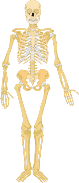
Skeleton Clip Art at Clker.com - vector clip art online, royalty free & public domain
Label Parts of the Human Eye - University of Dayton Parts of the Eye Select the correct label for each part of the eye. The image is taken from above the left eye. Click on the Score button to see how you did. Incorrect answers will be marked in red.
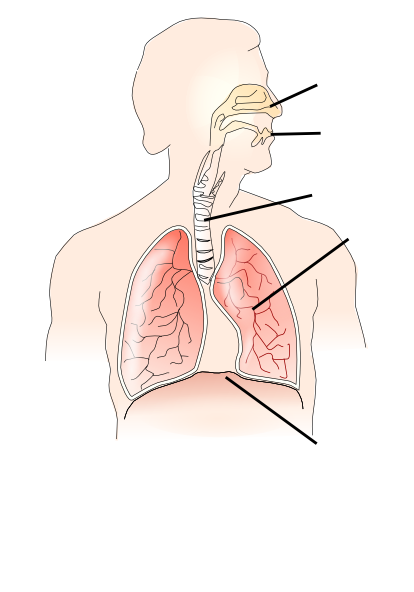
Unlabelled Respiratory System Clip Art at Clker.com - vector clip art online, royalty free ...
Eye Diagram - Differentiated Worksheets and EASEL Activities - Pinterest Description Use these simple eye diagrams to help students learn about the human eye. Three differentiated worksheets are included: 1. Write the words using a word bank 2. Cut and paste the words 3. Write the words without a word bank Labels include: eyebrow, eyelid, eyelashes, pupil, iris, and sclera.

BRAIN - SAGITTAL SECTION
Label Parts of the Human Ear - University of Dayton Label Parts of the Human Ear. Select One Auditory Canal Cochlea Cochlear Nerve Eustachian Tube Incus Malleus Oval Window Pinna Round Window Semicircular Canals Stapes Tympanic Membrane Vestibular Nerve. Select One Auditory Canal Cochlea Cochlear Nerve Eustachian Tube Incus Malleus Oval Window Pinna Round Window Semicircular Canals Stapes ...

Iran's 'eye for an eye' acid punishment postponed after outcry | Al Bawaba
PDF Eye Anatomy Handout - National Eye Institute of light entering the eye. Lens: The lens is a clear part of the eye behind the iris that helps to focus light, or an image, on the retina. Macula: The macula is the small, sensitive area of the retina that gives central vision. It is located in the center of the retina. Optic nerve: The optic nerve is the largest sensory nerve of the eye.
Post a Comment for "38 the human eye without labels"