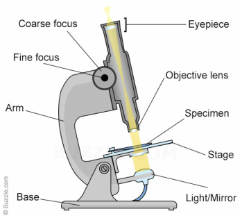
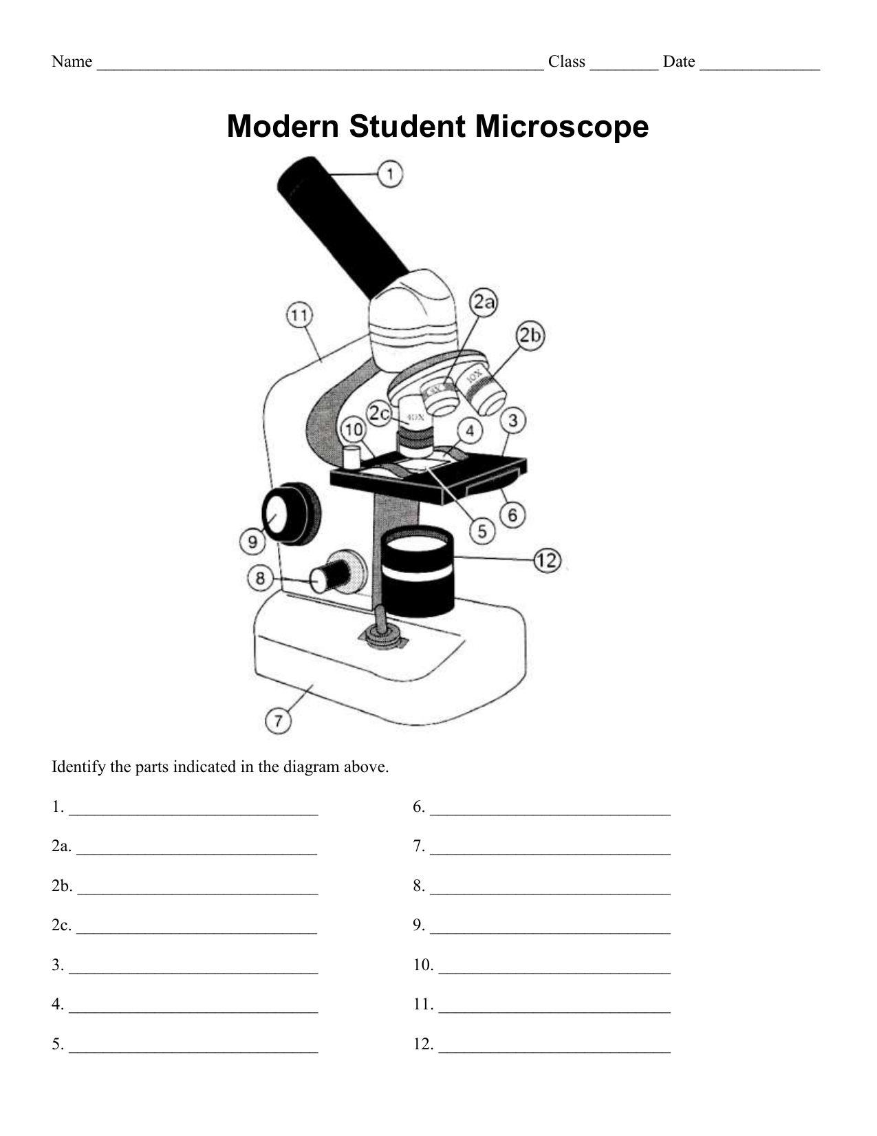
38 microscope diagram without labels
pages.zeiss.com › rs › 896-XMS-794Principles of Fluorescence and Fluorescence Microscopy - ZEISS fies the principle of the fluorescence microscope — without the light-filtering abilities of the purple glass window and the glass of white wine, Stokes would not have been able to observe any fluorescence at all. Using Stokes’ observation and the green fluorescent protein (GFP) as examples, this article will explain Electron microscope - Wikipedia An electron microscope is a microscope that uses a beam of accelerated electrons as a source of illumination. As the wavelength of an electron can be up to 100,000 times shorter than that of visible light photons, electron microscopes have a higher resolving power than light microscopes and can reveal the structure of smaller objects.. Electron microscopes use shaped magnetic …
Optical Sensor - an overview | ScienceDirect Topics 10.4.1 Sensors in cellular environments. Optical sensors operating in cellular environments can provide information about cell functions by probing molecules secreted from cells in situ and in real time without perturbing the cells. The optical properties of SWCNTs make them appealing for use in sensors under cellular environments because their emission is less likely to be absorbed …

Microscope diagram without labels
› books › NBK26880Looking at the Structure of Cells in the Microscope ... A special sample holder is used to keep this hydrated specimen at -160°C in the vacuum of the microscope, where it can be viewed directly without fixation, staining, or drying. Unlike negative staining, in which what is seen is the envelope of stain exclusion around the particle, hydrated cryoelectron microscopy produces an image from the ... Fluorescence Resonance Energy Transfer (FRET) Microscopy Microscope configurational parameters for fluorescence resonance energy transfer investigations vary with the requirements of the fluorophores, specimen, and imaging mode(s), but virtually any upright or inverted microscope can be retrofitted for FRET microscopy (see Figure 7). In general, the microscope should be equipped with a high-resolution (12-bit) cooled and … Fluorescence - Wikipedia Fluorescence is the emission of light by a substance that has absorbed light or other electromagnetic radiation.It is a form of luminescence.In most cases, the emitted light has a longer wavelength, and therefore a lower photon energy, than the absorbed radiation.A perceptible example of fluorescence occurs when the absorbed radiation is in the ultraviolet …
Microscope diagram without labels. Looking at the Structure of Cells in the Microscope A special sample holder is used to keep this hydrated specimen at -160°C in the vacuum of the microscope, where it can be viewed directly without fixation, staining, or drying. Unlike negative staining, in which what is seen is the envelope of stain exclusion around the particle, hydrated cryoelectron microscopy produces an image from the macromolecular structure itself. Rule based access control advantages and disadvantages Subjects and objects have clearances and labels, respectively, such as confidential, secret, and top secret. A subject may access an object only if the subject's clearance is equal to or greater than the object's label. Jul 25, 2019 · Role-Based Access Control (RBAC) is used by most OpenStack services to control user access to resources. Authorization is granted if a user has … › confocal-microscopes › lsm-980LSM 980 with Airyscan 2 – Confocal Microscope with Multiplex ... This requires excellent imaging performance combined with low phototoxicity and high speed. LSM 980, your platform for confocal 4D imaging, is optimized for simultaneous spectral detection of multiple weak labels with the highest light efficiency. Employ a wealth of fluorescent labels from 380 nm to 900 nm. en.wikipedia.org › wiki › Electron_microscopeElectron microscope - Wikipedia An electron microscope is a microscope that uses a beam of accelerated electrons as a source of illumination. As the wavelength of an electron can be up to 100,000 times shorter than that of visible light photons, electron microscopes have a higher resolving power than light microscopes and can reveal the structure of smaller objects.
LSM 980 with Airyscan 2 – Confocal Microscope with Multiplex To capture the complex world of biology, the ability to expand the number of labels is a great advantage. LSM 980 can image multiple labels simultaneously, covering a wide emission range up to 900 nm. These Cos-7 cells were labelled with 4 different fluorophores, two of which have their emission peak in the near infrared range (NIR), Alexa 700 ... en.wikipedia.org › wiki › FluorescenceFluorescence - Wikipedia Fluorescence is the emission of light by a substance that has absorbed light or other electromagnetic radiation.It is a form of luminescence.In most cases, the emitted light has a longer wavelength, and therefore a lower photon energy, than the absorbed radiation. › en › microscopeFluorescence Resonance Energy Transfer (FRET) Microscopy Presented in Figure 3 is a Jablonski diagram illustrating the coupled transitions involved between the donor emission and acceptor absorbance in fluorescence resonance energy transfer. Absorption and emission transitions are represented by straight vertical arrows (green and red, respectively), while vibrational relaxation is indicated by wavy ... › products › microscopeMicroscope Objective Lens | Products | Leica Microsystems The objective lens is a critical part of the microscope optics. The microscope objective is positioned near the sample, specimen, or object being observed. It has a very important role in imaging, as it forms the first magnified image of the sample. The numerical aperture (NA) of the objective indicates its ability to gather light and largely determines the microscope’s resolution, the ...
Microscope Objective Lens | Products | Leica Microsystems Microscope Objectives. Leica Microsystems – The Ultimate in Optical Competence. For more than 170 years Leica Microsystems has designed and produced top-class objectives for a wide variety of applications in research, industry and medicine. The optics specialists at Leica Microsystems bring the highest level of experience and expertise to bear in reducing … Principles of Fluorescence and Fluorescence Microscopy - ZEISS fies the principle of the fluorescence microscope — without the light-filtering abilities of the purple glass window and the glass of white wine, Stokes would not have been able to observe any fluorescence at all. Using Stokes’ observation and the green fluorescent protein (GFP) as examples, this article will explain fluorescence and fluorescence microscopy. The Principle of … An Overview of Hyphae in Fungi, Their Function and Types. 10.01.2022 · In this alternative exercise, students will create a 3D annotated diagram of hyphae and its stages and types using food items or any other … Fluorescence - Wikipedia Fluorescence is the emission of light by a substance that has absorbed light or other electromagnetic radiation.It is a form of luminescence.In most cases, the emitted light has a longer wavelength, and therefore a lower photon energy, than the absorbed radiation.A perceptible example of fluorescence occurs when the absorbed radiation is in the ultraviolet …
30 Label Diagram Of Microscope
Fluorescence Resonance Energy Transfer (FRET) Microscopy Microscope configurational parameters for fluorescence resonance energy transfer investigations vary with the requirements of the fluorophores, specimen, and imaging mode(s), but virtually any upright or inverted microscope can be retrofitted for FRET microscopy (see Figure 7). In general, the microscope should be equipped with a high-resolution (12-bit) cooled and …
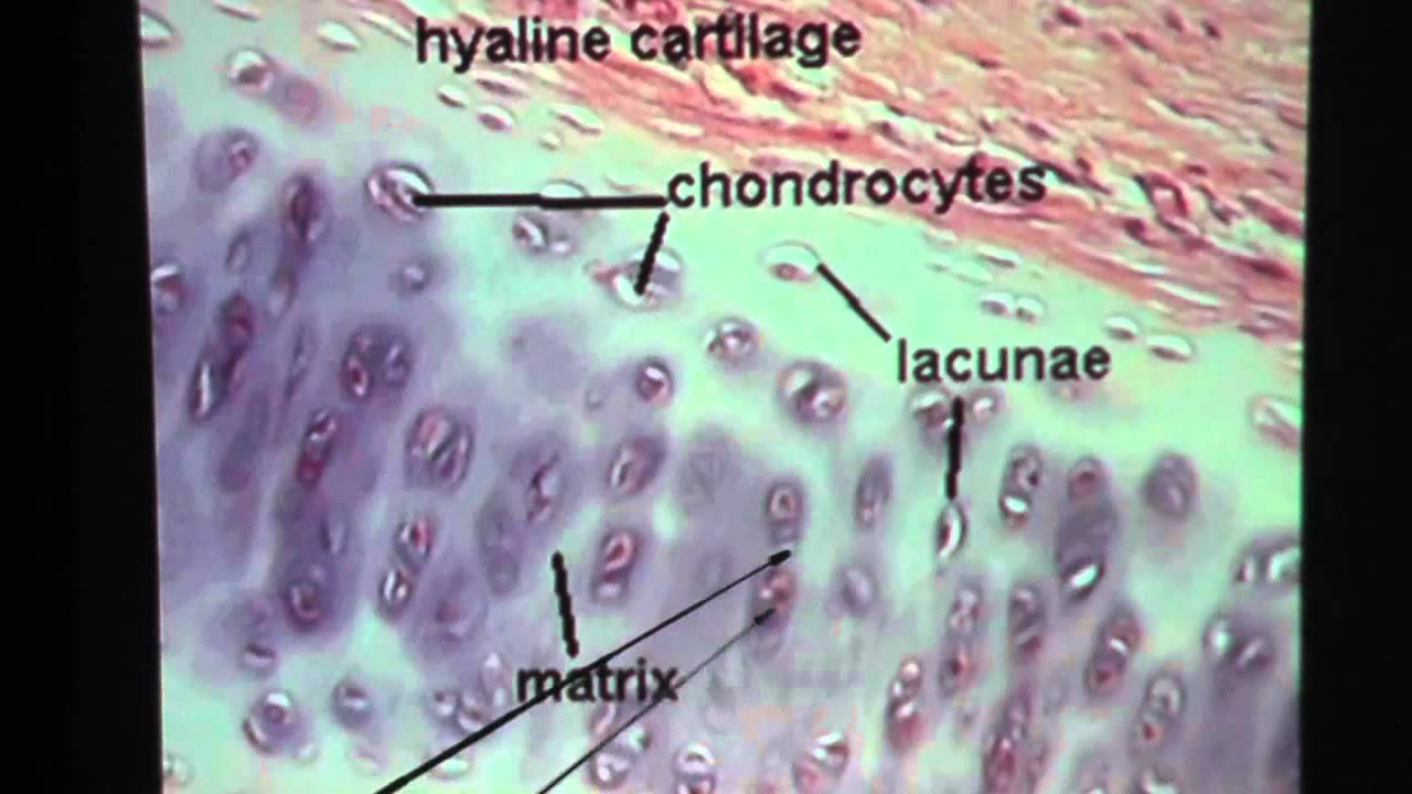
Hyaline Cartilage Connective Tissue - YouTube
› books › NBK26880Looking at the Structure of Cells in the Microscope ... A special sample holder is used to keep this hydrated specimen at -160°C in the vacuum of the microscope, where it can be viewed directly without fixation, staining, or drying. Unlike negative staining, in which what is seen is the envelope of stain exclusion around the particle, hydrated cryoelectron microscopy produces an image from the ...
Labeled-microscope-diagram

Label the diagram of the microscope and explain the role of each part: - Brainly.in
Guide to Using a Microscope - Year 8 Portfolio

A schematic diagram of the light path in the ptychographic microscope... | Download Scientific ...
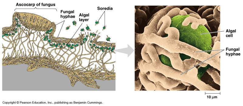
Fungi

31 Label The Indicated Parts Of The Microscope - Labels For Your Ideas

The Neuron Nerve Cell Nervous system, Brain PNG | PNGWave

Simple Microscope Labelled Diagram - Micropedia

Mr. Mak's Grade 8 Science Website

A Study of the Microscope and its Functions With a Labeled Diagram | Teaching ideas

Biology: January 2011
Microscope labeling, modern and classical types

Prokaryotic cells (Prokaryotes): Definition, Structure, Parts, Examples and Diagram

BIOLOGY ORDINARY LEVEL NOTES: CELL STRUCTURES
Search in gallery
Post a Comment for "38 microscope diagram without labels"