39 images of compound microscope with labels
Microscope Labeled Pictures, Images and Stock Photos Browse 49 microscope labeled stock photos and images available, or start a new search to explore more stock photos and images. Newest results Fluorescent Imaging immunofluorescence of cancer cells growing... Microscope diagram vector illustration. Labeled zoom instrument... Microscope diagram vector illustration. Compound Microscope with labels Stock Vector | Adobe Stock Download Compound Microscope with labels Stock Vector and explore similar vectors at Adobe Stock. Adobe Stock Photos Illustrations Vectors Videos Audio Templates Free Premium Editorial Fonts
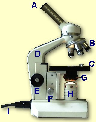
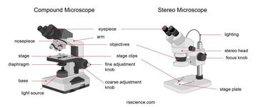
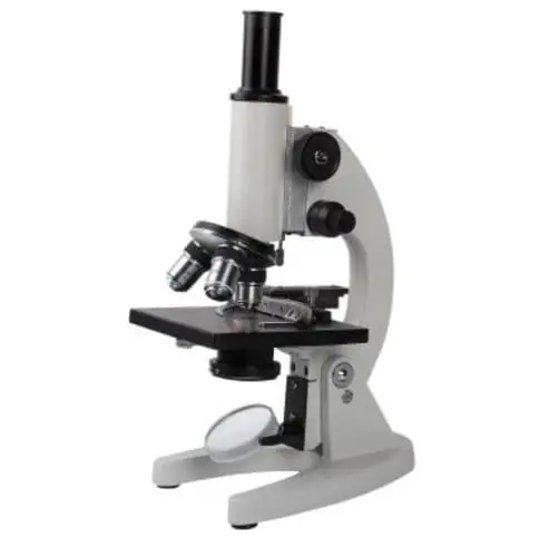
Microscope Parts and Functions First, the purpose of a microscope is to magnify a small object or to magnify the fine details of a larger object in order to examine minute specimens that cannot be seen by the naked eye. Here are the important compound microscope parts... Eyepiece: The lens the viewer looks through to see the specimen.

Images of compound microscope with labels
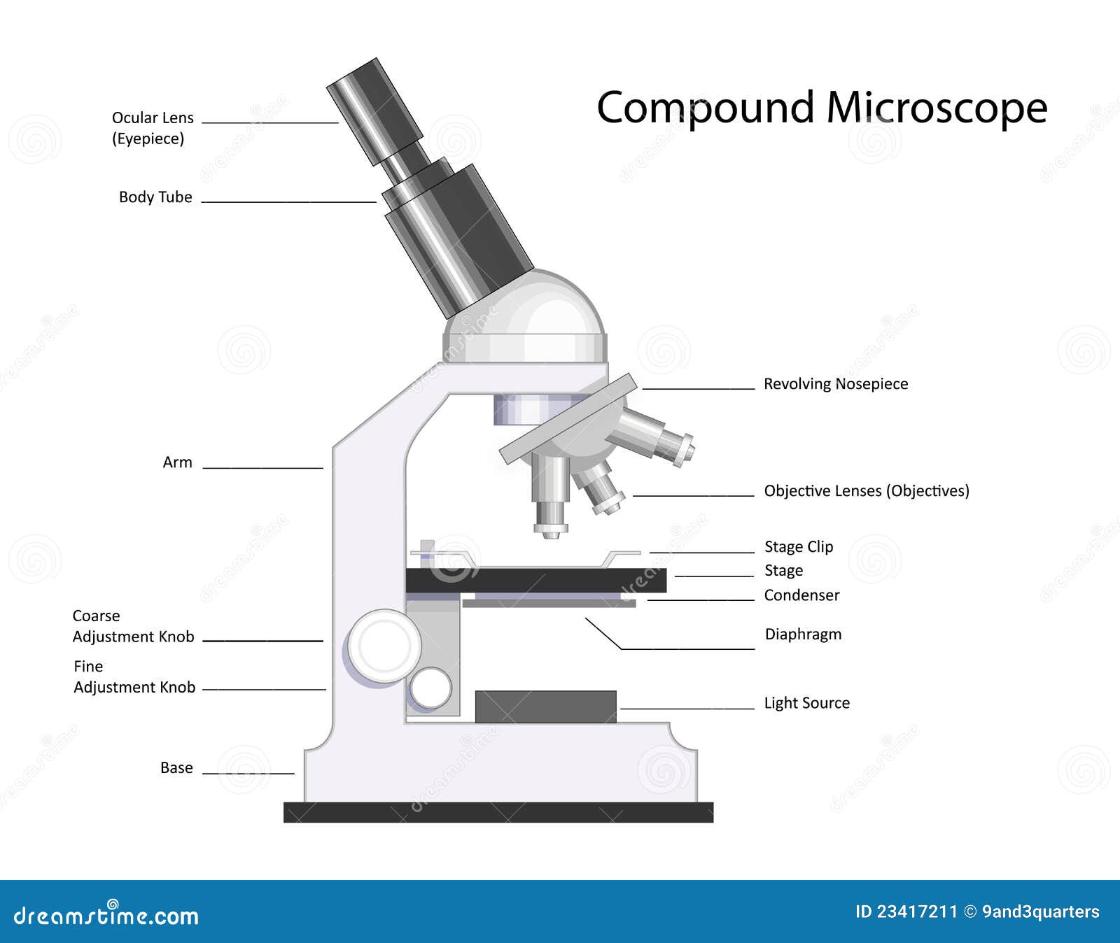
Electron microscope - Wikipedia An electron microscope is a microscope that uses a beam of accelerated electrons as a source of illumination. As the wavelength of an electron can be up to 100,000 times shorter than that of visible light photons , electron microscopes have a higher resolving power than light microscopes and can reveal the structure of smaller objects. What is a Compound Microscope? - Microscope Clarity A compound microscope utilizes a system of compounding lenses that enables the microscope to produce highly magnified images. Some of the lenses involved in this compound lens structure are the condenser lens, objective lens (which are themselves made up of several lenses), and the eyepiece lens. Compound microscopes can produce images magnified anywhere from 40x - 2,500x. 16 Parts of a Compound Microscope: Diagrams and Video In compound microscopes with two eye pieces there are prisms contained in the body that will also split the beam of light to enable you to view the image through both eye pieces. 2. Arm The arm of the microscope is another structural piece. The arm connects the base of the microscope to the head/body of the microscope.
Images of compound microscope with labels. Compound Microscope - Types, Parts, Diagram, Functions and Uses It comes with a wide body and base. Its distinct parts include a condenser, illumination, focus lock, mechanical stage, and a revolving nosepiece which can hold up to five objectives. It usually has a binocular head, which makes long-term observation easy. Image 22: An example of a research compound microscope. Inorganic Chemistry 4th edition, Catherine Housecroft Purple acid phosphatases (PAPs) are a group of metallohydrolases that contain a dinuclear Fe(III)M(II) center (M(II) = Fe, Mn, Zn) in the active site and are able to catalyze the hydrolysis of a variety of phosphoric acid esters. Label the microscope — Science Learning Hub All microscopes share features in common. In this interactive, you can label the different parts of a microscope. Use this with the Microscope parts activity to help students identify and label the main parts of a microscope and then describe their functions.. Drag and drop the text labels onto the microscope diagram. If you want to redo an answer, click on the box and the answer will go back ... Compound Microscope Stock Photos and Images - Alamy Find the perfect compound microscope stock photo. Huge collection, amazing choice, 100+ million high quality, affordable RF and RM images. ... Compound Microscope Stock Photos and Images (1,984) compound microscope isolated. Related searches: ... Method of illuminating compound microscope with gas lamp. Labels: C, ...
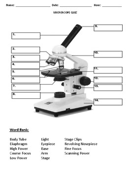
compound microscope parts (labeling) Flashcards | Quizlet what is 4? 40x objective lens - the "high" power objective lens with the most magnification. what is 5? stage clips - hold the slide in place on the stage. what is 6? iris diaphragm - controls the amount of light in the picture (the contrast) what is 7? illumination system - light source of the microscope. what is 8? Compound Light Microscope: Everything You Need to Know A compound light microscope is a type of light microscope that uses a compound lens system, meaning, it operates through two sets of lenses to magnify the image of a specimen. It's an upright microscope that produces a two-dimensional image and has a higher magnification than a stereoscopic microscope. It also goes by a couple of other names ... What is a Compound Microscope? - Study.com The body of the compound light microscope is the main part of the microscope, not to include the lights, focusing block, or stand of the microscope. The objective lenses and eyepiece are a part of ... Microscope Types (with labeled diagrams) and Functions A compound microscope: Is used to view samples that are not visible to the naked eye Uses two types of lenses - Objective and ocular lenses Has a higher level of magnification - Typically up to 2000x Is used in hospitals and forensic labs by scientists, biologists and researchers to study micro organisms Compound microscope labeled diagram
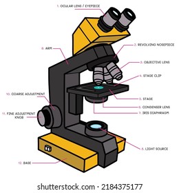
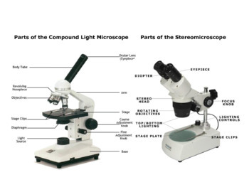
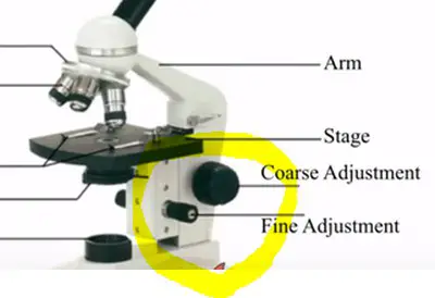
Parts of the Microscope with Labeling (also Free Printouts) Parts of the Microscope with Labeling (also Free Printouts) By Editorial Team March 7, 2022 A microscope is one of the invaluable tools in the laboratory setting. It is used to observe things that cannot be seen by the naked eye. Table of Contents 1. Eyepiece 2. Body tube/Head 3. Turret/Nose piece 4. Objective lenses 5. Knobs (fine and coarse) 6. Compound Microscope - Diagram (Parts labelled), Principle and Uses See: Labeled Diagram showing differences between compound and simple microscope parts Structural Components The three structural components include 1. Head This is the upper part of the microscope that houses the optical parts 2. Arm This part connects the head with the base and provides stability to the microscope. Parts of Stereo Microscope (Dissecting microscope) – labeled … Compared to a compound microscope where the objectives attached to the nosepiece can be seen and identified individually (based on color bands and their respective labels), the objectives of a dissecting microscope are located in a cylindrical cone and, therefore, are not directly seen. For the stereo microscope that comes with multiple objective lens sets (fixed power style), the … Drawing Of A Microscope And Label - Warehouse of Ideas Here presented 54+ microscope drawing and label images for free to download, print or share. Title Is Informative, Centered, And Larger Than Other Text. How to draw a microscope and label. Compound microscopes have furthered medical research, helped to solve crimes, and they have repeatedly proven invaluable in unlocking the secrets of the.
Microscope
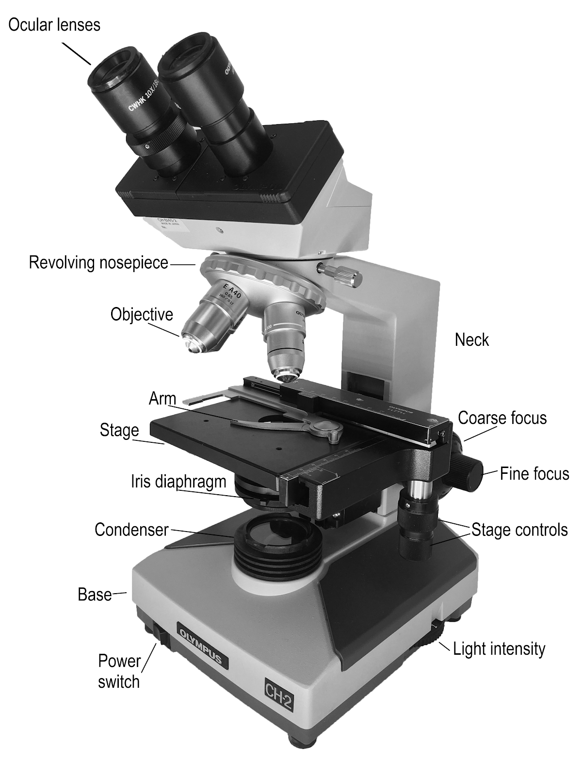
Compound Microscope Parts, Functions, and Labeled Diagram Compound Microscope Definitions for Labels. Eyepiece (ocular lens) with or without Pointer: The part that is looked through at the top of the compound microscope. Eyepieces typically have a magnification between 5x & 30x. Monocular or Binocular Head: Structural support that holds & connects the eyepieces to the objective lenses.
Compound Microscope Stock Illustrations – 734 Compound ...
What is Electron Microscopy? - UMASS Medical School Because of its great depth of focus, a scanning electron microscope is the EM analog of a stereo light microscope. It provides detailed images of the surfaces of cells and whole organisms that are not possible by TEM. It can also be used for particle counting and size determination, and for process control. It is termed a scanning electron microscope because the image is formed by …
Label Parts Of A Compound Microscope Teaching Resources | TpT
Looking at the Structure of Cells in the Microscope ... A typical animal cell is 10–20 μm in diameter, which is about one-fifth the size of the smallest particle visible to the naked eye. It was not until good light microscopes became available in the early part of the nineteenth century that all plant and animal tissues were discovered to be aggregates of individual cells.
The Compound Light Microscope Label the following parts on ...
A survey on deep learning in medical image analysis 01/12/2017 · A variety of image generation and enhancement methods using deep architectures have been proposed, ranging from removing obstructing elements in images, normalizing images, improving image quality, data completion, and pattern discovery. In image generation, 2D or 3D CNNs are used to convert one input image into another. Typically, these ...

Care and Structure of the Compound Microscope 1.jpg - Care ...
What is a Compound Microscope? - New York Microscope Company A compound microscope is an instrument that is used to view magnified images of small specimens on a glass slide. It can achieve higher levels of magnification than stereo or other low power microscopes and reduce chromatic aberration. It achieves this through the use of two or more lenses in the objective and the eyepiece.

Compound Microscope Parts
Diagram of a Compound Microscope - Biology Discussion Magnification of the Image of the Object by Compound Microscope: A bright-field or compound microscope is primarily used to enlarge or magnify the image of the object that is being viewed, which can not otherwise be seen by the naked eye. Magnification may be defined as the degree of enlargement of the image of an object provided by the microscope.

Parts of Compound Microscope | Botany
Parts of a microscope with functions and labeled diagram - Microbe Notes Q. Differentiate between a condenser and an Abbe condenser. Ans. Condensers are lenses that are used to collect and focus light from the illuminator into the specimen. They are found under the stage next to the diaphragm of the microscope. They play a major role in ensuring clear sharp images are produced with a high magnification of 400X and above.
3,343 Compound microscope Images, Stock Photos & Vectors ...
Solved Label the image of a compound light microscope using - Chegg Expert Answer. 100% (17 ratings) Transcribed image text: Label the image of a compound light microscope using the terms provided.
Parts of Stereo Microscope (Dissecting microscope) – labeled ...
Robert Hooke - Biography, Facts and Pictures Robert Hooke’s own illustration of his compound microscope, with labels added by this website. Hooke used his microscope to observe the smallest, previously hidden details of the natural world. His book Micrographia revealed and described his discoveries. Some people disputed his diagrams because they refused to believe what they showed. The world Hooke had discovered …

Parts of a Compound Microscope and Their Functions
(PDF) Introduction to Microscopy - ResearchGate Nov 08, 2017 · • In compound microscope it will be i.e 10 X, f= 16 mm; 40 X, f= 4 mm; 100 X, f= 1.8 mm. • Image produced by objective lens falls on the eyepiece lens serve as objec t. • Image formed in the ...
3,343 Compound microscope Images, Stock Photos & Vectors ...
300+ Free Microscope & Laboratory Images - Pixabay 399 Free images of Microscope. Related Images: laboratory science bacteria research scientist lab biology chemistry medical. Find your perfect microscope image. Free pictures to download and use in your next project.
3,343 Compound microscope Images, Stock Photos & Vectors ...
Food Calorimetry: How to Measure Calories in Food We have the compound microscope you are looking for! Digital Microscopes . Digital microscopes are great for large classroom computer combined instruction. Students can take images, videos, and more. Stereomicroscopes. Stereomicroscopes show 3D images vs. flat images and are easier to focus and use. They are great for first tme student use. Physical & Earth Sciences …
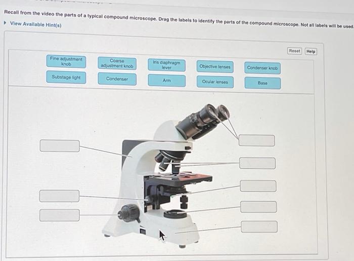
Solved Recall from the video the parts of a typical compound ...
Quantum Confinement Effect - an overview | ScienceDirect Topics In Nanostructured Semiconductor Oxides for the Next Generation of Electronics and Functional Devices, 2014. 6.5.2 PbS and PbSe quantum dot layers. It has been reported that the quantum confinement effect contributes to the extension of the photovoltaic potential of low-bandgap semiconductors such as PbS or PbSe (bandgaps are about 0.41 157 and 0.27 eV 158 for PbS …
Stereo & Compound Microscope Print Out by CalmAndConfidence | TpT
Labeling the Parts of the Microscope | Microscope World Resources Labeling the Parts of the Microscope. This activity has been designed for use in homes and schools. Each microscope layout (both blank and the version with answers) are available as PDF downloads. You can view a more in-depth review of each part of the microscope here.
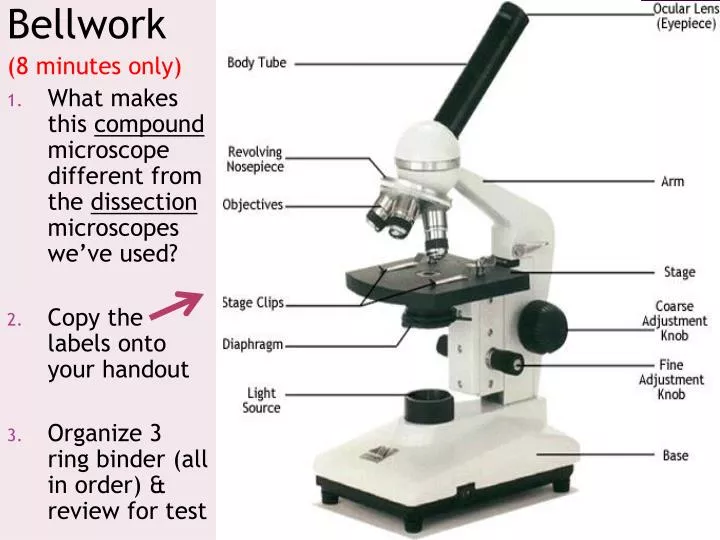
PPT - Bellwork (8 minutes only) PowerPoint Presentation, free ...
Compound Microscope- Definition, Labeled Diagram, Principle, Parts, Uses In order to ascertain the total magnification when viewing an image with a compound light microscope, take the power of the objective lens which is at 4x, 10x or 40x and multiply it by the power of the eyepiece which is typically 10x. Therefore, a 10x eyepiece used with a 40X objective lens will produce a magnification of 400X.
Label the Parts of a Compound Light microscope - BIOLOGY JUNCTION
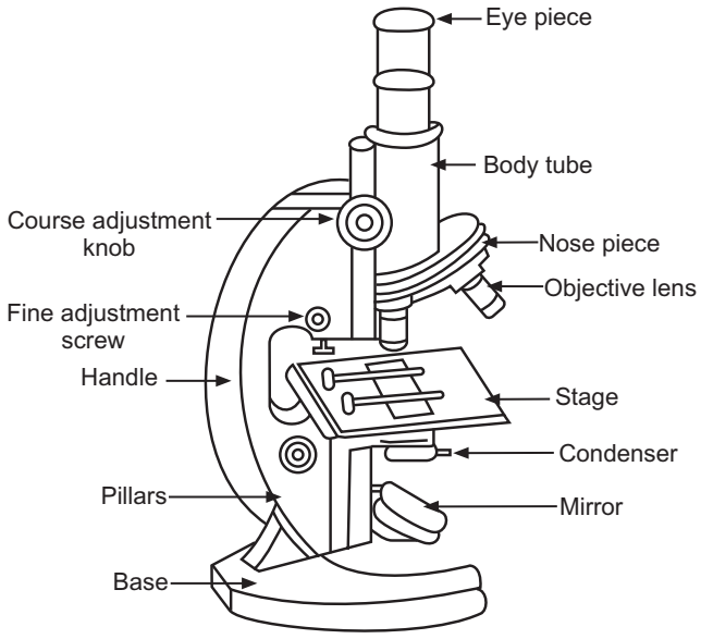
Labelled Diagram of Compound Microscope The below mentioned article provides a labelled diagram of compound microscope. Part # 1. The Stand: The stand is made up of a heavy foot which carries a curved inclinable limb or arm bearing the body tube. The foot is generally horse shoe-shaped structure (Fig. 2) which rests on table top or any other surface on which the microscope in kept.

label microscope diagram | Charts | Microscope, Anatomy bones ...
A general design of caging-group-free photoactivatable ... - Nature 21/07/2022 · The design of photoactivatable fluorophores—which are required for some super-resolution fluorescence microscopy methods—usually relies on light-sensitive protecting groups imparting ...

Compound Microscope Diagram | Quizlet
Compound Microscope: Definition, Diagram, Parts, Uses, Working ... - BYJUS A microscope with a high resolution and uses two sets of lenses providing a 2-dimensional image of the sample. The term compound refers to the usage of more than one lens in the microscope. Also, the compound microscope is one of the types of optical microscopes. The other type of optical microscope is a simple microscope.

3,343 Compound microscope Images, Stock Photos & Vectors ...
Histology Meyer's Histology - Online Interactive Atlas and Virtual Microscope Improve your identification and understanding of histological structures! View over over 4000 high resolution images of histological structures accompanied by interactive descriptive text that labels relevant histological details of every cell and tissue in the human body.

3,343 Compound microscope Images, Stock Photos & Vectors ...
Compound Microscope Illustrations & Vectors - Dreamstime New users enjoy 60% OFF. 192,670,685 stock photos online. Download 730 Compound Microscope Stock Illustrations, Vectors & Clipart for FREE or amazingly low rates! New users enjoy 60% OFF. 192,670,685 stock photos online. ... Compound Microscope. Clearly labeled vector of modern compound microscope. EPS 8 with no gradients or effects, layers ...

Compound Microscope Parts – Labeled Diagram and their ...
dock8 deficiency attenuates microglia colonization in early 17/08/2022 · B Representative images and quantification of 4 dpf apoeb WISH signals of dock8-2bp mutants, dock8-15,+5bp/-2bp compound mutants and siblings. Group sizes were at least n = 30 zebrafish embryos.

General Biology | Carlson Stock Art | General biology ...
Compound Microscope Labeled Diagram | Quizlet Compound Microscope Labeled + − Flashcards Learn Test Match Created by meganplocher734 Terms in this set (14) Eyepiece/Ocular lens Contains the ocular lens Body tube A hollow cylinder that holds the eyepiece. Arm Part that supports the microscope. Stage Supports the slide or specimen Coarse adjustment Knob

Compound Microscope – Diagram (Parts labelled), Principle and ...
Compound Microscope Parts - Labeled Diagram and their Functions Basically, compound microscopes generate magnified images through an aligned pair of the objective lens and the ocular lens. In contrast, "simple microscopes" have only one convex lens and function more like glass magnifiers. [In this figure] Two "antique" microscopes played significant roles in the history of biology.

Microscope Diagram and Functions | Microscope parts, Science ...
16 Parts of a Compound Microscope: Diagrams and Video In compound microscopes with two eye pieces there are prisms contained in the body that will also split the beam of light to enable you to view the image through both eye pieces. 2. Arm The arm of the microscope is another structural piece. The arm connects the base of the microscope to the head/body of the microscope.

13 parts of the Compound Light Microscope Diagram | Quizlet
What is a Compound Microscope? - Microscope Clarity A compound microscope utilizes a system of compounding lenses that enables the microscope to produce highly magnified images. Some of the lenses involved in this compound lens structure are the condenser lens, objective lens (which are themselves made up of several lenses), and the eyepiece lens. Compound microscopes can produce images magnified anywhere from 40x - 2,500x.

Compound Microscope Parts, Diagram Definition, Application ...
Electron microscope - Wikipedia An electron microscope is a microscope that uses a beam of accelerated electrons as a source of illumination. As the wavelength of an electron can be up to 100,000 times shorter than that of visible light photons , electron microscopes have a higher resolving power than light microscopes and can reveal the structure of smaller objects.
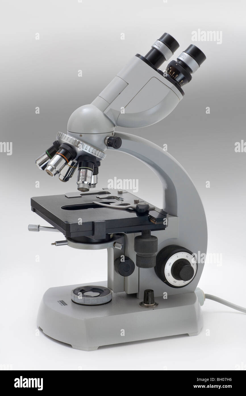
Compound microscope hi-res stock photography and images - Alamy

This is a common compound microscope. Label its parts from A ...

Compound Microscope: Parts of Compound Microscope

Solved Microscope parts/labeling 9 Label the image of a ...

Label Microscope Diagram - EnchantedLearning.com
Compound Microscope - Types, Parts, Diagram, Functions and ...

OMAX 40X-2500X Trinocular Biological Compound Microscope with Replaceable LED Light
Parts of the Microscope with Labeling (also Free Printouts ...
9.1: Using Microscopes - Biology LibreTexts

This is a common compound microscope Label its parts class 11 ...

The Microscope
Study of Compound Microscope - Solution Pharmacy
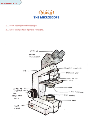
Microscope Activity - MICROBIOLOGY - 1... Draw a compound ...
Post a Comment for "39 images of compound microscope with labels"